Civil War Amputation
Procedures
Stephen
Smith, M.D. Handbook of Operative Surgery, 1863
Edited
by Dr. Michael Echols
From from the medical textbook
Handbook of Surgical Operations,
U. S. A. Medical Department, 1863,
(in this collection) written during the Civil War by
Stephen Smith,
M.D.,
with various drawings from the
medical literature. Drawings from Bourgery & Jocob
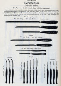
AMPUTATIONS IN GENERAL
Amputations are
performed through the shafts of bones, or through the joints; the
former are said to be in the continuity, and the latter,
in the contiguity.
Amputations In the Continuity:
There are several methods of shaping the external parts to cover the
stump in this operation :
Drawings from Bourgery & Jacob
ORIGINAL PHOTOS OF INSTRUMENTS AND PROCEDURES FROM
SMITH'S HAND- BOOK
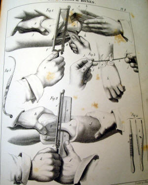
Circular Method.—There are
three principal steps in this operation:
1. Incision of the skin; 2.
Incision of the muscles; 3. Section of the bone.
To incise the skin easily and
neatly, the operator should stand upon the right side of the limb,
the left foot thrown forward and placed firmly upon the floor, the
right knee bending sufficiently to give freedom of motion to the
body; the left hand grasps the limb -above the point of operation,
and the handle of the knife is taken between the thumb and
forefinger of the right hand, being lightly supported by the other
fingers; stooping sufficiently to allow the right arm to encircle
the limb readily, he carries the knife around until the blade is
nearly perpendicular to the long axis of the limb on the side next
to him with the point downwards, and the hand of the operator above
the limb; he now commences the incision with the heel of the knife,
giving slightly sawing motions, and brings the hand under the limb,
and then directly upwards upon the side next to the operator, until
the heel touches the point of commencement; the handle of the knife
held thus delicately will change its relative positions as it passes
around the limb without the slightest embarrassment to the operator;
if the handle is firmly grasped in the whole hand, the incision
cannot be completed without the aid of the other hand, or an awkward
movement of the hand holding the knife; the ease with which the
incision is completed will depend much upon whether it commences
well down upon the side of the limb next to the operator. The skin
is raised from the first layer of muscles by dissection, and drawn
upwards, two or three inches, according to the diameter of the limb,
like the cuff of a coat.
2. The first layer of muscles is
divided at the margin of the retracted integument, in the same
manner as the incision of the skin is executed; this layer
is raised with the knife, and drawn still further upwards; and the
last layer of muscles is divided down to the bone by the
same sweep of the knife as before given. 3. The bone is then sawn at
the apex of the cone.
Modifications.—The chief
modifications are:—Louis divided the skin and superficial layer of
muscles at one incision, drew them up and divided the remainder;
Petit incised the skin, drew it up an inch, and then divided the
muscles directly to the bone; Alanson dissected up the skin and then
cut the muscles completely to the bone; Bell followed Alanson-, but
concluded by detaching the muscles from the bone, an inch or more,
with the point of a knife.
Flaps: Flaps may be
anterior, posterior, or lateral; they may be made from without
inwards, or from within outwards.
Single Flaps.—The operator
grasps the tissues on the anterior part of the limb, with the left
hand above the point of operation, and placing the heel of the knife
at the point of the fingers on the opposite side of the limb, with a
slight downward curve, he brings it over to the point of the thumb
on the opposite side, with one stroke dividing the tissues to the
bone; he now withdraws the knife until the point rests in the angle
of the wound, when he thrusts it under the bone, taking care that
the point emerges at the angle of the wound on the opposite side
where the incision commenced ; he now makes a flap from the
posterior part of the limb of sufficient length to cover the stump;
the muscles are dissected from the bone with the amputating knife or
a scalpel; the operation is very rapid, the knife not being raised
from the limb.
Double Flaps: The operator grasps the tissues on the upper part of the limb with
the left hand, the thumb and fingers resting at the middle of the
limb on opposite sides; the knife, a, is then entered at the
thumb and thrust through above the bone, emerging on the opposite
side at the point where the fingers rest, and passed downwards and
outwards, c, making an anterior flap, b, of the
required length; it is again re-entered at the same point, and
passing beneath the bone emerges from the same point on the opposite
side, and a flap is made from the posterior part of the limb; both
flaps are forcibly retracted, the muscles dissected from the bone,
and the bone divided.
Modifications. Ravaton
divided the soft parts circularly down to the bone, and then made
two lateral incisions to the requisite extent and raised
quadrilateral flaps; Sedillot first made two small flaps, turned
them back, and completed the operation as in the circular method;
Langenbeck made the flaps from without inwards.
Rectangular Flaps. First mark on the limb with ink two flaps; the size of the long
flap is determined by the circumference of the limb at the place of
amputation, its length and its width being each equal to half the
circumference. The long flap is therefore a perfect square, and is
long enough to pull easily over the end of the bone. In selecting
the structures for its formation, such parts must be taken as do not
contain the larger blood-vessels and nerves. The flap so formed will
be for the most part anterior in position as far as regards the
general aspect of the body, but superior when the patient is in the
recumbent posture, as during the after treatment. The short flap,
containing the chief vessels and veins, is in length one- fourth of
the other. The flaps being formed, the bone sawn, and the arteries
tied, the long flap is folded over the end of the bone; each of its
free angles is then fixed by suture to the corresponding free angles
of the short flap. One or two more sutures complete the transverse
line of union of the flaps. At each side the short flap is united to
the corresponding portion of the long one by a point of suture, and
one suture more unites the reflected portion of the long flap to its unreflected (Fig. 95) portion. Thus the transverse of the union is
bounded at each end by a short lateral line at right angles to it.
Oval Method.—In this
operation the incision may be made like an inverted V, the apex
being a little below the point where the bone is to be sawn; the
incision being extended quite down to the bone before the lower
portion of the flap containing the large vessels is divided; or a
perfect oval may be marked out by the first incision, and
the operation completed as in the former case. The oval method is
seldom adopted except in amputation at the joints.
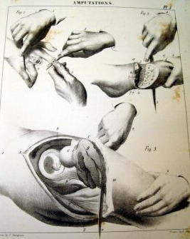
Amputations In the
Contiguity.—The methods of amputation already described may be
employed in the disarticulation of limbs. The instruments required
are a single knife, having a narrow blade, to enable it to move more
readily over the irregularities of the joint, with a thick back to
give it 'firmness, and an artery forceps. The chief points to be
well considered by the
operator are thus concisely stated by
Malgaigne (Operative Surgery,
Am. Edition):
The operator has here three
special objects in view: 1. To well recognise the articulation
before commencing. 2. The flesh being divided, to traverse the
articulation without hesitation, destroying all its means of
attachment. 3. To preserve flesh and integument enough.'
Whence the following rules:
To Recognize the Articulation:
The surgeon should have the disposition of the articulation so
fixed in his mind, that he could, without having it under his eyes,
trace it out exactly. In this way, recognizing one part of the
articulation, he is sure of the others, neither the blood nor the
soft parts causing the knife to deviate. He must also know the
direction of the ligaments, to attack them more surely; their
length, to cut them between their attachments; their breadth, to
divide them completely.
1. The surest guides in finding
the joints are the osseous projections. It is with them you must
first occupy yourself. To find them, you may place the limb in a
position that causes them to project; seek them on the side where
they are most prominent; put aside by careful pressure the soft
parts, fat, or oedema that mask their projections; and, lastly, seek
them by starting from a
known point: for example, passing the finger along the shank of a
long bone till you reach its extremity.
2. The second indication
consists in the folds of the skin, sometimes placed immediately
above the joint, sometimes some way from it.
3. You may, as a third resource,
cause to be prominent to the sight and touch the tendons which are
inserted near the joint. To effect this, cause the muscles to
contract; this is usually sufficient: or you may render their
projection greater by opposing the movement of the limb which their
contraction tends to execute.
4. If all these trials fail,
you may assist yourself by the neighboring tuberosities, whether-or
not they be in the same line, provided their distance and connexions
are well determined beforehand. It is objected that the relations,
and especially the distance, vary in different subjects;
consequently, they can only give us a tolerably close indication:
but certainly there is never more than some lines difference; and it
is better to have such approximation than none at all.
5. If these means do not
suffice, seize the limb with the right hand, and seek the joint with
the left, moving the limb slightly, and thus try to mark out the two
diameters of the joint, or, in other terms, the point of entrance
and exit of the knife.
6. Lastly, supposing that all
these indications do not afford a certain result, incise the skin in
the most suitable direction, and, after having raised it, assure
yourself by the touch of the articular line. If the touch does not
point it out, place the knife in the angle of the wound nearest
yourself, its heel perpendicular to the horizon, and the edge
perpendicular to the bone, and thus move it along the bone with a
sawing sidelong movement, without taking it off, and the pressure
will cause the knife to enter the joint when it reaches it.
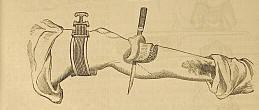
Use of
the tourniquet, Liston amputation knife & capital saw
To Traverse the Articulation:
1. The articulation recognised,
or at all events presumed to be so, as we have directed, the index
finger and thumb should rest applied on the two extremities of the
articular diameter until the knife replaces them. If this search has
been made with the right hand, substitute the left hand for it
before seizing the knife. In this way you mark the point of entrance
and exit of the knife.
2. If you attack the joint by
its dorsal surface, semiflex the limb, to extend the parts and
enlarge the articular line. Without this precaution, you often fall
on the neighboring joint, as happens on the foot and hand.
3. The knife should not
generally be carried into the joint without having first cut its
principal means of connexion, which should be divided from without
inwards.
4. In joints with several
projections and inter lockings, commence by the internal or external
side. As the knife opens one joint, do not push it in there, but go
on dividing and opening further. In this way, the
ligaments are not put out of the reach of the knife, or shielded
by bony projections.
5. An important fact. An
articulation, that offers to the anatomist a surface equal to
one inch, presents to the operator at least four. So long as the
ligaments are divided between their attachments, it is of slight
importance whether during their division the knife fall on the articular line or at the side of it.
6. The dorsal and lateral
ligaments being cut, we can generally engage all the blade of
the knife between the articular surfaces; but if there are
interosseous ligaments, they must be first divided. Carry the
point of the knife directly on them: as they are divided, the
joint opens.
7. To destroy these
ligaments, you must know the interstices between the bones
through which they may best be attacked. In general, on the hand
and foot, the bones, very compact on their dorsal surface, leave
between them on their palmar and plantar surfaces intervals
which lodge these ligaments. Carry the knife under these
intervals, inclining the handle towards yourself, and making it
form an angle of forty-five degrees anteriorly; then raise it up
to a right angle. The ligaments divided by this movement allow
the articulation to be opened sufficiently for the knife to
enter it.
8. It is useless to luxate:
it strains the parts very painfully; and if you separate the
parts very much on one side, you apply those of the other
together. If in cases of difficulty you have recourse to this
means, luxate downwards as far as half the dorso- palmar
diameter, and then vice versd. But it is better to
separate the parts by slight traction parallel to the axis of
the stump: this ordinarily suffices. The heel and point of the
knife should always move in the same line. If, in bringing the
knife out of the joint, you dread jagging the integuments, push
them gently aside with your left forefinger and thumb.
To Preserve Sufficient
Flap:
1. The proceedings vary
according to the method, and often even in each method.
2. In the circular, you can
generally count only on the skin to cover the surface of the
wound. Make the incision at a sufficient distance from the
joint, and dissect back the skin as a cuff. If there are muscles
under it, you may cut them obliquely on the plan of Alanson, or
divide them perpendicularly on a level with the joint.
3. The oval method is
ordinarily performed by tracing on the dorsal surface a V
incision reversed, the ends of which are joined by a
semicircular incision round the palmar surface. When there are
any large vessels, leave them in the portion to be divided last,
as in the method by flaps, so as to be able to compress the
artery before dividing it beyond the part compressed.
4. In most of the oval
proceedings, the second incision is made to join the first at
its point of commencement. A loss of substance is the
consequence; or, if the V terminates on a level with th«
articulation, there is considerable difficulty in getting the
knife to act in disarticulating. I lay down here as a general
rule, expose the joint to be destroyed by a longitudinal
incision passing half an inch, at least, above, and one inch
below it. The two branches of the V, which fall on the
inferior part of this incision, leave, as it were, two small
flaps at the-upper part, which do not hinder immediate and
linear union, and which perfectly cover the osseous prominences
left by the disarticulation.
5. The methods by one or
two flaps are executed in two ways. Sometimes the flaps are made
first, before touching the joint; but most usually a simple
incision is made first, or the least important flap, and the
second is not begun till after the disarticulation.
6. The knife having
traversed all the joint, when the bones are large and uneven, as
in the foot and hand, the instrument must be withdrawn, and its
point placed horizontally in the extremity of the joint next the
hand operating, and its way cut by pressing from right to left.
7. To avoid terminating the
flap by a point, the knife must be held horizontally close to
the bones, and kept so to the required extent, cutting
freely.
8. It is well, before you
terminate your flap, to apply it to the part to be covered, to
see if it is long enough.
9. If there remain any
tendons beyond the bleeding edge, cut them off with a scissors.
10. If you fear too much
retraction of the skin, do not divide it until the muscles have
retracted.
11. You may cut your flap
from engorged tissues, so long as the engorgement is not
malignant.
12. You may operate when
there is not enough skin to make a flap; a cicatrix will be
formed on the articular surfaces."
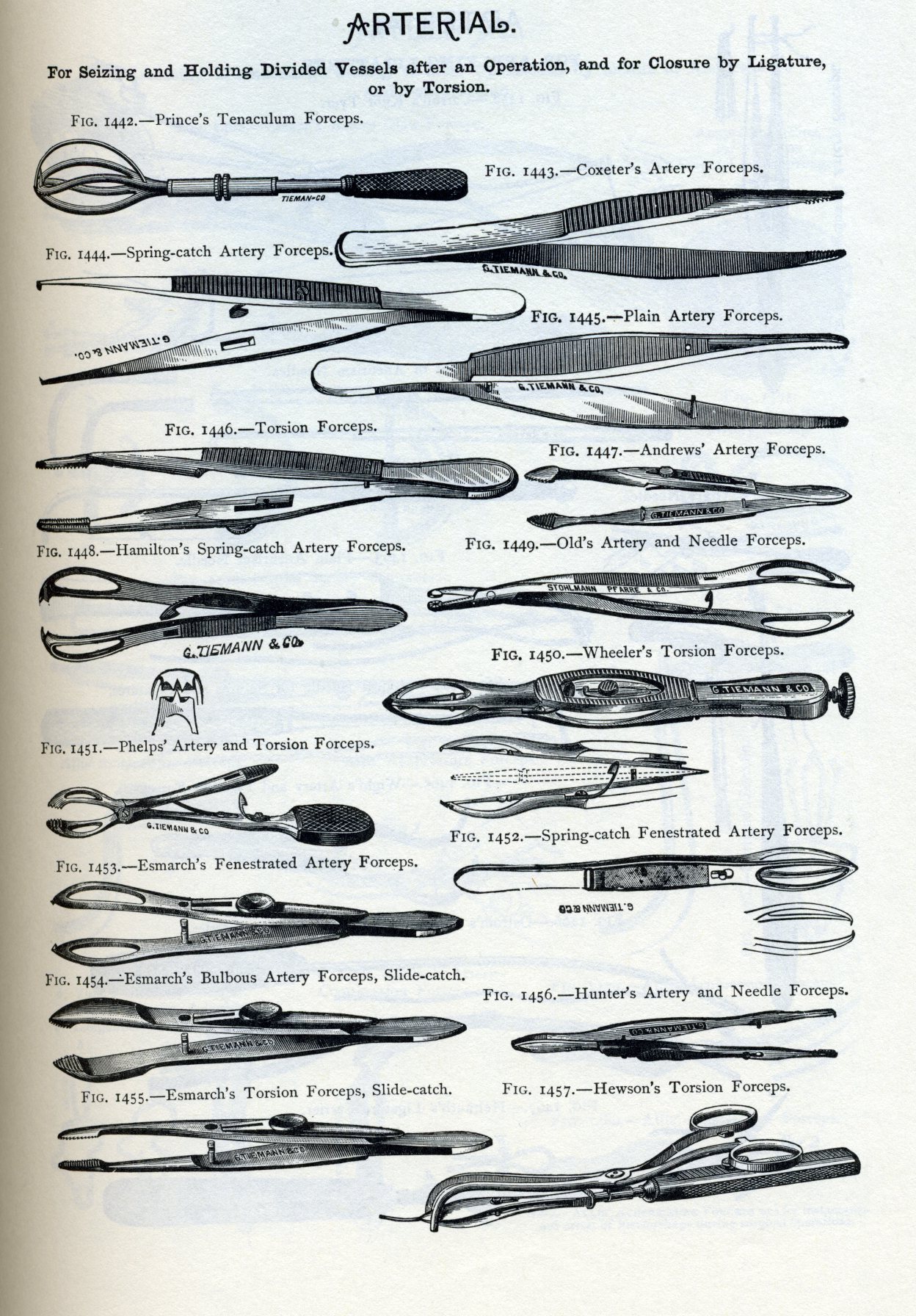
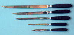
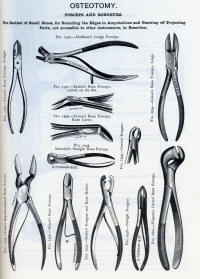
Instruments: The following
instruments are required to form a complete amputating case:—A
long and short knife, catling, metacarpal saw, scalpel,
tenaculum, saw, bone forceps, artery forceps, needles, and
tourniquet.
(Instrument drawings
are from
the 1880's Tiemann Catalog, other examples are from a
Civil War military Hospital Department surgical set by
Hernstein)
|
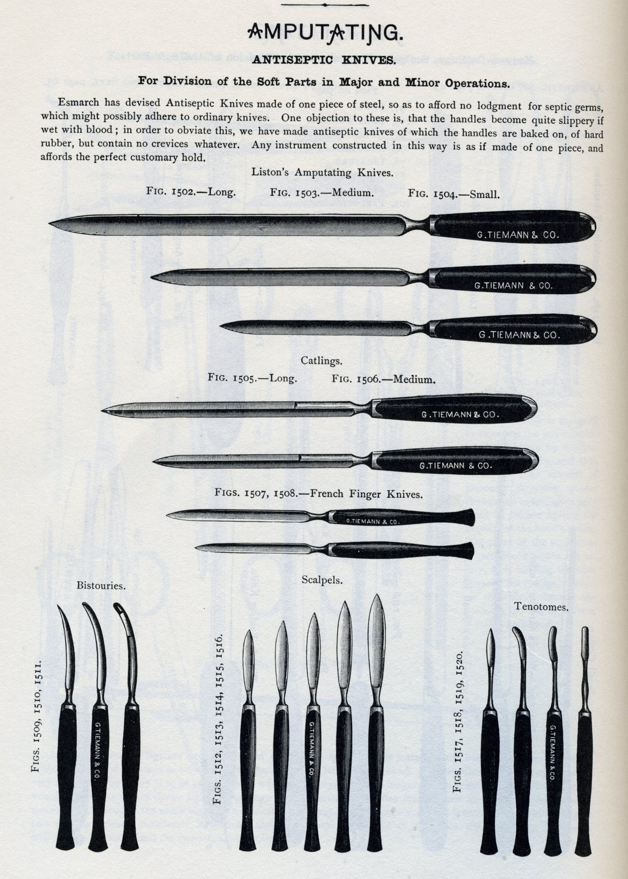
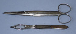
Knife, Catling, and
Scalpel: The knife, a, selected for each operation
should be of about twice the length of the diameter of the limb;
the catling, is a double-edged knife, the two edges being
parallel until they converge to form the point; the scalpel, is
large and strong, having a firm handle.
 |
 |
|
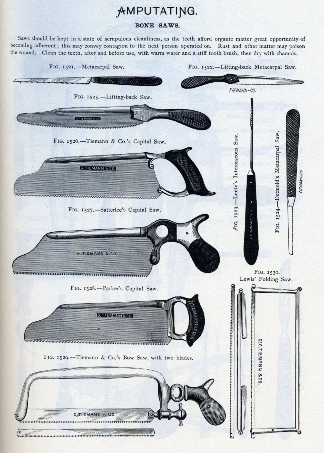
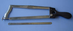
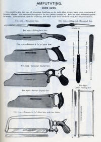
The Saw: The form of
saw generally used is represented in Dr. Wood's case; the edge
should be straight, the teeth fine,
and so set as never to allow the saw to bend in
its passage through the bone; a saw having fine teeth is but
little liable to lacerate the periosteum, and produces a
comparatively small amount of comminution of the osseous tissue.
 |
 |
|
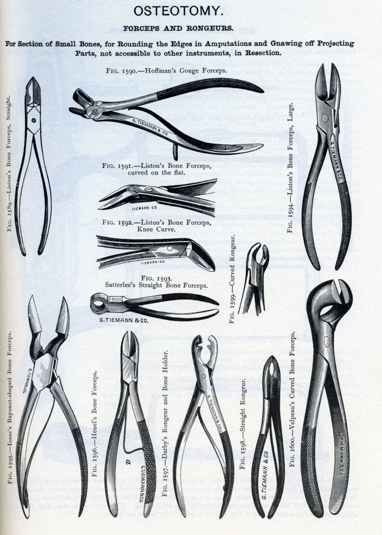
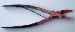
Bone Forceps: The
forceps in common use is that of Liston.
 |
 |
|
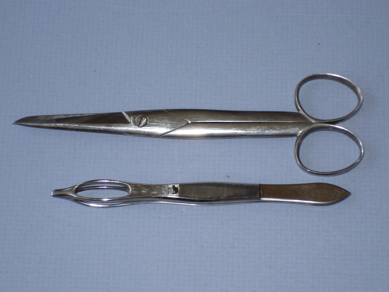
In addition to the common
artery forceps a pair of small forceps will
sometimes be found useful in dissecting out vessels.
 |
 |
|
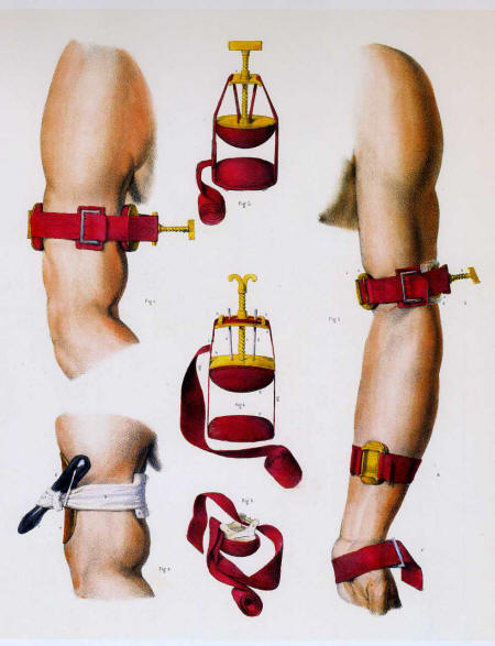
Tourniquet
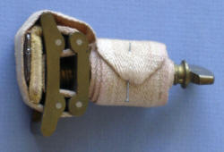 |
 |
Arrangements.—The retractor
is a piece of muslin, half a yard long, and half an inch wide,
split in the centre, half its length; it is used by passing the
tails on either side of the bone, and the extremities being
seized by an assistant, the flaps are forcibly drawn upwards.
The sponges should be fine, and in large number. These
preliminaries being arranged, the patient is placed in a
recumbent position, on a table of such height as to enable the
operator to manipulate with his instruments about the limb with
perfect freedom ; if it is an amputation by the circular method
the limb should be much more elevated than if the flap operation
is performed. The anaesthetic is administered by an assistant,
and when the patient is under its influence a second assistant
applies the tourniquet as
directed,
page 26, but as far as possible from the point of operation;
previously to its being tightened, if it is desirable to save to
the system the largest possible amount of blood, the limb is
elevated and rubbed towards the heart to force the blood in the
superficial veins beyond the point of amputation; compression of
the artery by the fingers of an assistant should never be relied
on when a tourniquet can be used. A third assistant should hold
and steady the extremity of the limb, and a fourth is in waiting
to retract the flaps, either with his hands or with the
retractor, and to apply ligatures; a fifth uses the sponges. It
is usual to have an assistant to hand the instruments to the
operator, but it is much better and more convenient for the
operator to place the instruments which he will require on a low
bench by his side, in the order of the steps of the operation;
an assistant frequently mistakes the instrument called for, and
by his confusion often delays vexatiously an important step of
the operation. The operator takes a position, in general, upon
the right side of the limb. In amputations of the leg, however,
the best position for sawing the bones is on the inside.
Operative Procedure.—An
amputation in the continuity involves the following steps:
Incision of Soft Parts.—-The
method being selected, the operator proceeds, according to the
rules already given, to form the coverings of the bone from the
soft parts; the bone having been exposed, an assistant, cither
with his hands or the retractor, draws the flaps firmly upwards
and maintains them in that situation.
Incision of the Bone.—The
periosteum having been divided completely around the bone, as
high up in the flap as possible, the saw is employed, in
imitation of the cabinet-maker, the heel being first applied,
and the saw drawn slowly but firmly across the bone to make a
groove in which it will work, and then moved with as much
rapidity as the operator may choose, until the bone is nearly
divided, when it is to be moved more slowly to avoid splintering
the last connexions. With the bone forceps any sharp or
projecting edges are clipped off, and the end of the bone
bevelled smoothly. Where there is a single bone it will be found
easier to apply the saw nearly perpendicularly on the side
opposite to the operator; where there are two bones the saw
should be first and last applied to the larger and firmer bone,
the smaller bone being completely divided while the saw is
engaged in the larger bone.
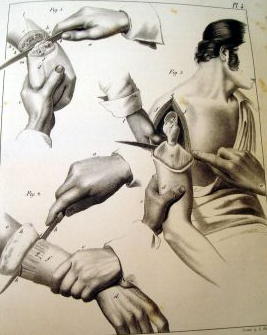
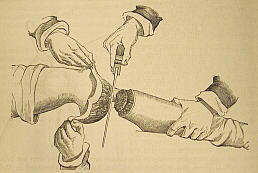
Ligating Arteries.—The
arteries should be tied according to the rules given,
page 31.
If a vein bleeds freely, a ligature should be applied. Before
the stump is dressed any dark clot on its face should be
removed, and a bleeding vessel sought for underneath it.
Dressings.—The flaps
should be brought accurately together, and maintained by silk or
metallic ligatures; the sutures should be taken deeply in the
lips of the wound, from a quarter to half an inch, and at
sufficient intervals to support the parts in apposition, with
the assistance of adhesive straps applied in the intervals. The
external dressings should be light and cool; the limb is then
placed in an elevated position, and protected from all pressure
and irritation.
FOR THE
ORIGINAL PHOTOS OF INSTRUMENTS AND PROCEDURES FROM
SMITH'S HAND- BOOK,
CLICK HERE
Article on suturing during the
Civil War
Article on
anesthesia during the Civil War
Article on ligation of an
artery during the Civil War
|